DREAMIES T-M: Infant Hearing Protection for MR Imaging.
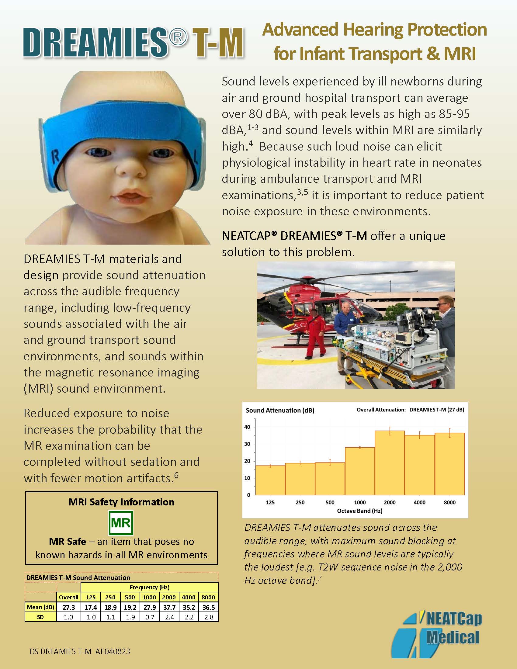
Reduced exposure to noise increases the probability that the MR examination can be completed without sedation and with fewer motion artifacts [M5].
DREAMIES T-M block noise across the full spectrum of sound, with maximum sound blocking at frequencies where MRI noise is the loudest [M7].
The most common methods used for obtaining infant MR imaging without sedation - patient immobilization and Feed-and-Swaddle – are often successful for exams of no more than 20-30 minutes in duration [M8].
Clinical users have reported improved results when using DREAMIES T-M with both methods. You may be able to reduce patient preparation time, and increase MR imaging study time by using DREAMIES T-M.

Clinical Results: DREAMIES T-M with papoose immobilizer
Results Include:
- Preemies prepped in papoose by nurse in NICU
- MRI scans not synchronized with feed
- No sedation
- Babies sleeping/quiet through entire MRI exam
- Good imaging – no repeat scans

Clinical Results DREAMIES T-M With Feed-And-Swaddle
Results Include:
- Feed-and-Swaddle conducted immediately before MRI scans
- No sedation
- Babies sleeping/quiet through entire MRI exam
- *Up to 60 minutes of good, complete Structural, Functional and Biochemical Imaging (T1-, T2-, PD-weighted imaging and EPI, DTI, SWI and DWI) vs. 20-30 min with foam ear plug/foam earmuff combination [E13]
What Nurses and Technologists are saying about using DREAMIES T-M in MRI
Jeanne
NICU Director“We feel that the MRI patient hearing is protected when using the DREAMIES T-M device compared to what we have used in the past. None of our patients using DREAMIES T-M needed to have an MRI repeated due to a poor study. The patients have slept though the MRI studies without the use of any sedation!”
Nancy
MR Imaging Technologist, Pediatric Research“We have used DREAMIES T-M during research MRI examinations on infants 3 days to 4 months of age. We don’t sedate – we “feed and bundle.” DREAMIES T-M have enabled us to continue scanning much longer, motion-free. Our subjects have stayed asleep for the entire research scan which includes DTI, BOLD, and ASL and other structural imaging sequences. This is extremely rare. DREAMIES T-M could be the game changer we have been waiting for!”
Noise Exposure and Sedation During MRI Examinations
Noise Exposure and Sedation During MRI Examinations
Magnetic resonance imaging (MRI) has become an important tool to augment brain ultrasound in the evaluation of neonates, especially in preterm and very low birth weight infants, due to its better sensitivity detection of for white matter lesions and brain abnormalities than cranial ultrasound [M1].
Obtaining high-quality MR images requires the patient to remain very still during the examination. So-called “motion artifacts” (blurred or poor image quality) that occur when the patient’s body moves may cause MR examinations to be rushed or terminated prematurely, requiring the examination to be repeated at a later time.
Loud noise generated by the MR scanner can elicit autonomic instability, as reflected in cardiac rhythm changes in neonates [M3, M4] and patient agitation resulting in motion artifacts in the scans.
For neonates and infants, it is generally recognized that some type of physical immobilization is required to obtain sufficient MR image quality. Additionally, sedation (or even anesthesia) while undesirable — sometimes is necessary to calm patients during MRI studies [M5]. However, sedation of neonates sufficient to ensure lack of movement can involve serious risks to patient respiratory stability [M9].
The sound levels during high-field-strength MR scanning are high. MR brain imaging pulse sequences average approximately 98 dBA for a conventional MRI scanner and approximately 88 dBA for a specially designed NICU scanner [M2]. Loud noise generated by the MR scanner is an important clinical issue in neonatal MRI examinations because such noise can elicit autonomic instability, as reflected in cardiac rhythm changes, in neonates [M3, M4].
For these reasons, passive hearing protectors (e.g., foam earplugs and/or ear muffs) capable of abating noise by 10 to 30 dB [M6] are used on patients, including infants and neonates [M2] in an attempt to reduce noise exposure during MRI examinations. Unfortunately, many such hearing protectors do not properly fit neonates and small infants so their effectiveness is limited.
DREAMIES T-M are compatible – and have been found to significantly improve results – with the two most commonly used non-sedative methods for infant immobilization during MRI: physical restraint, e.g. with a papoose board, and Feed-and-Swaddle.
Physical Restraint:
This method of infant immobilization relies upon a single Velcro strap to stabilize the infant patient’s head – and a papoose instead of cloth blankets to hold the infant’s body still. This method does not require the MR scanning time to be synchronized with the feeding schedule.
Feed-and-Swaddle:
The conventional “feed-and-swaddle” method (also known as feed-and-bundle or feed-and-wrap,) refers to the use of feeding and swaddling to induce natural sleep in infants. It requires close coordination between the MRI suite and the NICU or Pediatric In-patient Service to synchronize the time of PO feeding with the time of the MR exam. Unfortunately, the feed and swaddle technique is less reliable when applied to preemies and/or MR spinal examinations [M8] – and is not applicable to emergent situations or patients who are receiving continuous feeds.


